
Achilles Tendonitis vs. Achilles Tendon Tear:
How to Tell the Difference
Both conditions cause pain in the back of the leg — but one is a gradual overuse problem and the other is a surgical emergency. Knowing the difference could change the course of your recovery.

The Achilles tendon is the strongest tendon in the human body — and one of the most commonly injured. Whether it is the gradual, aching buildup of tendonitis that follows weeks of increased running mileage, or the sudden, devastating pop of a complete rupture during a pickup basketball game, Achilles injuries demand accurate diagnosis and timely treatment. Getting it wrong — minimizing a rupture as “just tendonitis,” or treating chronic tendinopathy without addressing its mechanical cause — leads to prolonged disability and, in the case of a missed rupture, potential permanent loss of function.
The Achilles Tendon: Anatomy and Function
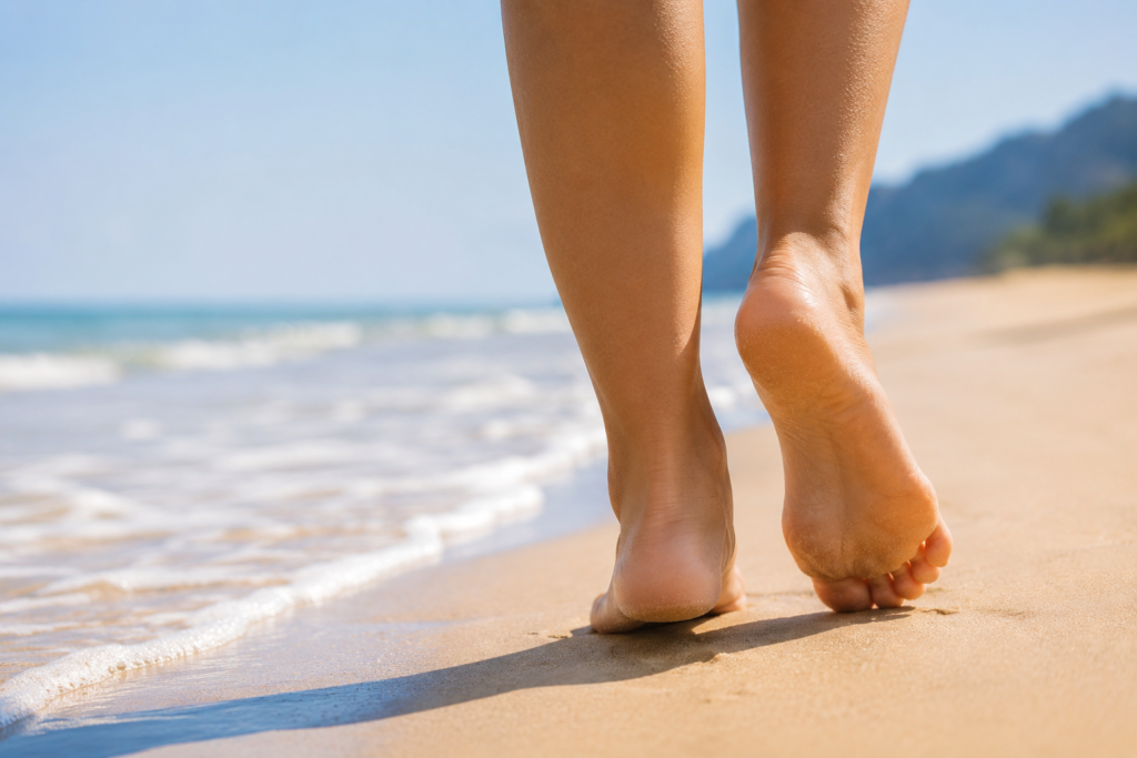
The Achilles tendon is the thick, cord-like structure running down the back of the lower leg, connecting the gastrocnemius and soleus muscles of the calf to the posterior calcaneus (heel bone). It is the largest and strongest tendon in the body, capable of withstanding forces of up to 8 times body weight during running and explosive movement.
Despite its strength, the Achilles tendon has a known vulnerability: a watershed zone of relatively poor blood supply located approximately 2 to 6 centimeters above the calcaneal insertion. This is precisely where the majority of both degenerative tendinopathy and acute ruptures occur. Reduced vascularity in this region means slower healing, impaired remodeling after microtears, and accumulation of degenerative change over time — the biological foundation of chronic Achilles pathology.
The tendon serves two critical mechanical functions: plantarflexion (pointing the foot downward) and energy storage and release during the propulsive phase of gait. When the Achilles is compromised — whether by tendinopathy or rupture — walking, running, and stair-climbing are all meaningfully affected.
Achilles Tendonitis and Tendinopathy — The Overuse Spectrum
The term “Achilles tendonitis” implies acute inflammation of the tendon — and while true inflammatory tendonitis does occur, particularly in early-stage presentations, the majority of what patients and clinicians call “Achilles tendonitis” is more accurately described as Achilles tendinopathy: a degenerative process within the tendon substance driven by repetitive mechanical overload that exceeds the tendon’s capacity to remodel and repair.
At the cellular level, tendinopathy involves disorganization of collagen fibers, neovascularization (ingrowth of abnormal blood vessels and nerve endings that contribute to pain), increased ground substance, and progressive loss of tensile strength. The tendon does not become inflamed in the traditional sense — it degenerates. This distinction matters clinically because true anti-inflammatory medications (NSAIDs, corticosteroids) have a limited role in tendinopathy beyond short-term symptom management, while mechanical loading through structured rehabilitation is the cornerstone of treatment.
Two Anatomically Distinct Presentations
Affects the tendon body 2–6 cm above the heel bone — the classic “watershed zone.” Pain, thickening, and nodularity in the mid-tendon. Most common in runners and active individuals. The most responsive to eccentric loading rehabilitation.
Affects the point where the tendon meets the heel bone. Often associated with a Haglund’s deformity (“pump bump”) — a bony prominence that impinges on the tendon. Pain at the back of the heel, worsened by shoe counter pressure. Less responsive to eccentric exercises; often requires modification of technique.
Symptoms of Achilles Tendonitis / Tendinopathy
The most common precipitating factor for Achilles tendinopathy is a rapid increase in training load — more mileage, more intensity, more hill work, or a return to activity after a period of rest — without adequate time for the tendon to adapt. The classic presentation is a runner who has increased weekly mileage quickly, or an occasional athlete (“weekend warrior”) whose Achilles has not been conditioned for the demands placed on it.
Other contributing factors include poor footwear, sudden changes in training surface, tight calf musculature, excessive pronation, and a cavus (high-arched) foot type that concentrates force on the posterior heel.
Achilles Tendon Tear — Partial and Complete Rupture
An Achilles tendon rupture is a partial or complete disruption of the tendon fibers — and it is one of the most commonly missed major injuries in orthopedic medicine. Studies consistently show that up to 25% of acute Achilles tendon ruptures are missed or significantly delayed in diagnosis because the patient can still walk, the swelling is variable, and the dramatic pop they describe does not match what they — or sometimes their initial provider — expect a serious injury to look like.
Ruptures occur most commonly in the watershed zone 2 to 6 centimeters above the calcaneal insertion — the same region affected by tendinopathy — and in many cases occur in tendons that were already degeneratively weakened, even if asymptomatic. This is why ruptures so frequently occur during seemingly routine activities rather than extreme athletic exertion.
Partial vs. Complete Rupture
Some tendon fibers remain intact. The patient may retain some plantarflexion strength. Diagnosis is more challenging — MRI is often required to quantify the extent of tearing. Treatment ranges from functional bracing to surgery depending on the percentage of tendon involved and functional demands.
Full disruption of all tendon fibers. Classic presentation: sudden pop, acute pain, inability to rise on toes. A palpable gap may be felt in the tendon. Thompson test is positive (no plantarflexion with calf squeeze). Requires urgent evaluation — surgical or non-surgical treatment must begin promptly for best outcomes.
Classic Symptoms of an Achilles Tendon Rupture
One of the most dangerous misconceptions about Achilles ruptures is the assumption that if you can walk, the tendon must be intact. This is false. Other muscles — the peroneal muscles, the tibialis posterior, and the toe flexors — provide enough residual plantarflexion to allow a flat-footed gait even with a complete rupture. The key test is the single-leg heel rise: if you cannot rise up on your toes on the injured side, you need urgent evaluation regardless of your ability to walk. Delaying treatment beyond 2 weeks significantly worsens surgical outcomes.
Side-by-Side Comparison: How to Tell the Difference
| Feature | Achilles Tendinopathy | Achilles Tendon Rupture |
|---|---|---|
| Onset | Gradual — days to weeks of building pain | Sudden — immediate at moment of injury |
| Mechanism | Repetitive overload, training error | Single explosive force — push-off, jump, sprint |
| Pain character | Dull, aching; stiff in the morning; improves with warm-up | Sharp, severe; immediate; may lessen but weakness persists |
| Audible/felt pop | No | Yes — very characteristic of complete rupture |
| Ability to rise on toes | Usually preserved, though painful | Lost on single-leg test — key clinical sign |
| Palpable tendon gap | No — tendon thickened but continuous | May be present with complete rupture |
| Tendon on exam | Tender, thickened, possibly nodular | Discontinuous; Thompson test positive |
| Bruising | Absent or minimal | Often present — spreads around ankle and heel |
| X-ray findings | Usually normal; may show calcification at insertion | Soft tissue shadow disruption; Kager’s triangle changes |
| MRI findings | Tendon thickening, signal changes, neovascularization | Partial or complete fiber discontinuity, hematoma |
| Urgency | Urgent but not emergent — evaluate within days to weeks | Urgent — evaluate within 24–72 hours for best outcomes |
The Thompson Test — A Simple Bedside Diagnosis
The Thompson Test: Diagnosing Rupture in 10 Seconds
The Thompson test (also called the Simmonds-Thompson test) is the most reliable bedside examination for Achilles tendon rupture — and it can be performed by any clinician in seconds. The patient lies prone (face down) on the examination table with both feet hanging over the edge. The examiner firmly squeezes the calf muscle of the affected leg with both hands.
Normal result: The foot plantarflexes (points downward) — indicating the tendon is intact and transmitting the force from the calf to the heel.
Positive result (rupture): The foot does not move, or moves significantly less than the uninjured side — indicating the tendon is no longer transmitting force from the calf to the calcaneus. A positive Thompson test in the context of acute injury is diagnostic of a complete Achilles tendon rupture until proven otherwise and requires urgent imaging and surgical consultation.
It is important to note that the Thompson test can occasionally yield a false-negative result — particularly with partial ruptures where some fibers remain intact, or in the early hours after injury before significant swelling has developed. Clinical suspicion should always drive the decision to obtain MRI when the diagnosis is uncertain, even if the Thompson test appears equivocal.
Who Is at Risk — and Why
Understanding the risk factors for Achilles injury helps both in prevention and in interpreting a patient’s history when arriving at a diagnosis.
Risk Factors for Achilles Tendinopathy
Risk Factors Specific to Achilles Rupture
Fluoroquinolone antibiotics — including ciprofloxacin (Cipro) and levofloxacin (Levaquin) — carry a well-documented black-box warning for tendon rupture, including the Achilles tendon. The risk is greatest in patients over 60, those taking concurrent corticosteroids, and those with renal impairment. If you are prescribed a fluoroquinolone and develop new Achilles pain during or after the course of antibiotics, seek evaluation promptly and do not push through the discomfort.
How a Podiatric Surgeon Diagnoses Achilles Injuries
When a patient presents to my office with Achilles pain — whether gradual or sudden — the evaluation follows a systematic approach designed to accurately classify the injury before any treatment decision is made.
History is the most revealing part of the evaluation. How did the pain start? Was there a pop? What activity were you doing? Have you had Achilles pain before? Are you taking any medications — particularly fluoroquinolones or steroids? How long has it been since the injury? A clear mechanism of sudden onset during explosive activity with an audible pop is strongly suggestive of rupture; a gradual buildup over weeks in a runner who recently increased mileage points toward tendinopathy.
Physical examination includes the Thompson test, single-leg heel rise assessment, palpation of the tendon along its entire length, assessment of the Haglund’s prominence at the insertion, range of motion testing, and gait analysis. The presence or absence of a palpable gap in the tendon and the response to the Thompson test are the two most diagnostically decisive findings.
Weight-bearing X-rays of the foot and ankle are obtained to evaluate for calcaneal bony pathology, Haglund’s deformity at the insertion, intratendinous calcification (a marker of chronic tendinopathy), and Kager’s triangle changes that may suggest rupture on plain film.
MRI is the gold standard imaging modality for Achilles pathology. It allows precise characterization of tendon thickness, internal signal changes consistent with tendinopathy, the extent and location of tearing in partial ruptures, the degree of tendon retraction in complete ruptures, and the condition of adjacent structures. MRI is particularly important when the clinical diagnosis is uncertain, when partial rupture needs to be distinguished from complete rupture, or when surgical planning requires detailed anatomical information.
Ultrasound is a useful dynamic imaging tool — it allows real-time assessment of the tendon during movement and can detect neovascularization within the degenerative tendon. It is operator-dependent and less reliable for precise characterization of rupture extent than MRI but offers the advantage of in-office assessment without radiation.
Treatment: Achilles Tendonitis and Tendinopathy
The treatment of Achilles tendinopathy is primarily non-surgical, and the vast majority of patients respond well to a structured, progressive rehabilitation program when it is followed consistently. The key word is consistently — Achilles tendinopathy is notorious for slow, non-linear recovery, and patients who abandon rehabilitation during good periods invariably experience setbacks.
Conservative Treatment — The Foundation
The Alfredson eccentric heel drop protocol has the strongest evidence base of any conservative treatment for non-insertional Achilles tendinopathy. The exercise involves standing on the edge of a step on the ball of the foot, rising up on both feet, then lowering slowly on the affected foot alone — working the tendon through a loaded lengthening contraction. Performed twice daily at 3 sets of 15 repetitions, with progressive loading added as tolerated over 12 weeks.
For insertional tendinopathy, the exercise is modified to avoid the painful end-range dorsiflexion — the heel drop is performed on flat ground rather than off a step.
Interventional Options When Conservative Care Is Insufficient
Platelet-rich plasma (PRP) injection — autologous growth factors drawn from the patient’s own blood and injected into the degenerative tendon zone. Evidence for PRP in Achilles tendinopathy is accumulating, with several randomized controlled trials showing benefit in chronic non-insertional tendinopathy. PRP is not a substitute for rehabilitation — it is an adjunct that may accelerate biological repair when combined with loading exercise.
High-volume injection (HVI) — injection of saline and corticosteroid around the tendon to strip neovascular ingrowth and interrupt the abnormal nerve supply contributing to pain. Used selectively in refractory cases under ultrasound guidance.
Extracorporeal shockwave therapy (ESWT) — focused acoustic energy delivered to the degenerative tendon to stimulate biologic repair. Supported by level I evidence for chronic insertional and non-insertional Achilles tendinopathy that has not responded to 3 months of conservative care.
Surgical debridement — for patients who have failed 6 or more months of structured conservative treatment, surgical removal of the degenerative tendon tissue and any associated bone spurs (particularly at the insertion) provides reliable pain relief in approximately 80 to 85% of cases.
Treatment: Achilles Tendon Rupture
Management of an acute Achilles tendon rupture involves a choice between two evidence-based approaches: surgical repair and non-surgical functional bracing with accelerated rehabilitation. Both have demonstrated good outcomes in appropriately selected patients, and the decision should be individualized based on patient age, activity level, medical comorbidities, and surgeon experience.
Surgical Repair
Primary surgical repair involves end-to-end re-approximation of the ruptured tendon ends through an open or minimally invasive approach, secured with non-absorbable sutures. Surgery restores near-normal tendon anatomy, allows earlier rehabilitation, and produces lower re-rupture rates (approximately 3–5%) compared to non-surgical management (approximately 10–12% in older studies, lower with modern functional bracing protocols).
Surgical repair is generally preferred for:
Non-Surgical Functional Bracing
Modern non-surgical management uses a functional bracing protocol — an equinus cast or CAM boot that holds the foot in plantarflexion to allow the tendon ends to approximate — followed by a structured, accelerated rehabilitation program with progressive weight-bearing. When executed correctly, this approach produces excellent functional outcomes comparable to surgery in appropriate candidates, without the risks of wound complications, infection, or nerve injury.
Non-surgical management is generally preferred for:
Regardless of whether surgery or bracing is chosen, prompt initiation of treatment is essential. With complete rupture, the tendon ends begin to retract and fibrous tissue fills the gap within days of injury. Delays beyond 2 to 4 weeks increase the difficulty of surgical repair, the length of the gap requiring bridging, and the likelihood of a weaker repair. If you suspect an Achilles rupture, do not wait for the swelling to go down — seek evaluation within 24 to 72 hours.
Recovery Timeline
After Achilles Tendon Repair Surgery
After Achilles Tendinopathy Rehabilitation
Recovery from Achilles tendinopathy is non-linear and highly individual. Mild, early-stage presentations may resolve in 6 to 8 weeks with consistent loading exercise and activity modification. Chronic tendinopathy that has persisted for months or years typically requires 3 to 6 months of structured rehabilitation before meaningful improvement is sustained. Patience and consistency are the most important determinants of outcome — patients who stop the program during pain-free periods almost always relapse.
Frequently Asked Questions
A strain or partial tear may be difficult to distinguish from a complete rupture on history alone — both can cause significant pain and swelling. The most reliable clinical differentiator is the single-leg heel rise test: if you can rise up on the toes of the injured foot, the tendon is at least partially functional. If you cannot, urgent evaluation is required. The Thompson test performed by a clinician is the most accurate bedside assessment. When in doubt, an MRI provides definitive characterization of the injury.
Ice is appropriate for acute injury, acute flares, and post-exercise symptom management — it reduces swelling and provides analgesia. For chronic Achilles tendinopathy between flares, heat before activity can help with tissue extensibility, while ice after activity reduces post-load soreness. Neither modality directly addresses the underlying tendon pathology — loading exercise is the only intervention that promotes tendon remodeling at the structural level.
In most cases, yes — with modification. Complete cessation of activity is rarely necessary and may actually slow recovery, as the tendon requires progressive loading to remodel. The general guideline is to reduce volume and intensity to a level where pain during and after running does not exceed 3 to 4 out of 10, and where pain returns to baseline within 24 hours of a run. If pain is consistently above this threshold or takes more than 24 hours to settle, loading is excessive and should be reduced. Running through severe pain risks progression to a partial tear.
Corticosteroid injection directly into the Achilles tendon is generally contraindicated due to the well-documented risk of tendon weakening and subsequent rupture. Injection around the tendon (peritendinous), performed under ultrasound guidance, is safer and may be used to treat paratendinitis (inflammation of the sheath surrounding the tendon) rather than the tendon body itself. For chronic Achilles tendinopathy, PRP and high-volume injection are preferred over corticosteroid. If you have been offered a direct Achilles tendon corticosteroid injection, please discuss the rupture risk with your provider before proceeding.
The honest answer is that a healed Achilles tendon is not identical to an uninjured one — repaired or conservatively managed tendons are typically larger in cross-section, have some scar tissue, and may have a mildly lower maximal force capacity than the contralateral side. However, the vast majority of patients — including competitive athletes — return to full sport and function at or very close to their pre-injury level. Return-to-sport rates after surgical repair are high. The most important factor in achieving a full recovery is commitment to structured rehabilitation through the entire 6 to 12 month process.
No — the relationship between Achilles tendinopathy and rupture is more complex than a simple progression. Many patients with tendinopathy never rupture, and many ruptures occur in tendons with no prior symptoms. However, tendinopathy does indicate degenerative weakening of the tendon — reduced collagen organization and tensile strength — which increases vulnerability to rupture under acute load. Patients with known Achilles tendinopathy should be counseled to avoid sudden, high-intensity activities (explosive sprinting, jumping) without adequate warm-up and conditioning, and to take new or worsening pain seriously rather than pushing through it.
Achilles Pain Deserves an Accurate Diagnosis
Whether it’s tendonitis that won’t resolve or an injury that happened suddenly — the right next step is a proper evaluation. Don’t wait to find out which one you’re dealing with.
Medical Disclaimer: The information in this article is for general educational purposes only and does not constitute individualized medical advice. Please consult a licensed podiatric physician for evaluation and treatment of any foot or ankle condition.
Explore Our Expert Care
Discover the achievements of Foot and Ankle Medical Group, where we have transformed lives through dedicated foot and ankle care. Take a look at some key statistics:
20+
Years of Expertise
200,000+
Successful Treatments
14,000+
Active Patients
5+
Dedicated Specialists
Begin Your Journey to Foot Health at Foot and Ankle Medical Group
At Foot and Ankle Medical Group, we prioritize your health above all. Schedule an appointment today and receive top-tier care from our team of skilled physicians. Committed to excellence, we employ advanced treatments and cutting-edge technology to enhance your foot and ankle health.